Major Instruments

Electrophoresis Unit

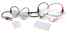
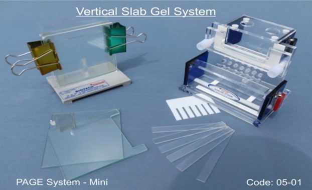
Electrophoresis describes particle mobility in a gel or fluid in a homogeneous electric field. Electrophoresis separates molecules by charge, size, and binding affinity. The method is used to isolate and study biomolecules such DNA, RNA, proteins, nucleic acids, plasmids, and their fragments. Paternity and forensic science employ electrophoresis to detect source DNA. Anaphoresis is anion electrophoresis. Cataphoresis is cation electrophoresis.

Real Time PCR

RT-PCR is a laboratory procedure that reverses RNA into DNA (called complementary DNA or cDNA) and amplifies particular DNA targets (PCR). It measures RNA amounts. Fluorescence-based real-time PCR or quantitative PCR monitors the amplification process (qPCR). In research and clinical contexts, combined RT-PCR and qPCR measure gene expression and viral RNA. RT-PCR and qPCR are synonymous due to their tight relationship. RT-PCR may be used without qPCR for molecular cloning, sequencing, or RNA detection, which may be confusing. qPCR may be performed without RT-PCR to count DNA copies.

Cooling Centrifuge

Centrifuges provide continual force to specimens to separate fluid components. Spinning the fluid at high speed in a container separates liquids from solids or cream from milk. It radiates denser particles. Lighter objects go to the centre. Radial acceleration in a laboratory centrifuge causes denser particles to sink. A centrifuge may filter fluid contaminants.

UV Trans illuminator

Molecular biology labs utilise UV-transilluminators to observe electrophoresed DNA or RNA in agarose gels. The agarose gel is coloured with a fluorescent nucleic acid-binding dye during or after electrophoresis. UVB light fluoresces DNA/dye in the stained gel. This method is used to size PCR products, purify DNA segments following restriction enzyme digests, quantify DNA, and validate RNA integrity after extraction. This Tutorial shows how to create a UVB (310nm) transilluminator with a 7 × 7 cm window for studying ethidium bromide (SYBR-Safe) stained DNA mini-gels. After collecting all supplies, assembly takes 1-2 hours. Needs soldering.

Magnetic Stirrer

A magnetic stirrer uses a rotating magnetic field from a rotating magnet or stationary electromagnet to spin a stirrer bar in a liquid to swiftly mix the solution. Richard Stringham of Utah patented the first magnetic stirrer in 1917. Modern magnetic stirrers use electric motors to spin the magnets. A magnetic stirring system usually has a connected liquid heating system. Magnetic stirrers can stir low-viscosity volumes. To avoid magnetic field interference, use stirrers with glass or non-metallic beakers. Stirring in academics, business, agriculture, health care, homebrewing, etc. is good.

Blotting Unit

Western blots identify protein molecules in a mixture. This combination can contain all tissue- or cell-specific proteins. Western blots can quantify protein expression and size. This approach was called because its resemblance to the Southern blot. Western blots begin by combining the protein sample with sodium dodecyl sulphate, which unfolds the proteins into linear chains and gives them a negative charge. Gel electrophoresis separates protein molecules by size. Proteins are transported from gel to blotting membrane after separation. Western blotting refers to the complete process, even if this phase is what gives it its name.